Is Coronary Artery Disease Hereditary? Genetic Links
January 24, 2025
3 mins
What if your doctor could produce clear images of your circulatory system’s complex pipework of interconnected blood vessels in a safe and simple way? Having this ability could lead to early diagnosis of blood vessel problems and a proactive approach to healthcare. As a diagnostic tool, it would help identify the precise location of a blood vessel blockage and lead to immediate intervention to limit major complications.
The technology described already exists. It’s called an angiogram and it is a commonly performed procedure designed to let doctors examine blood vessels with the aid of intravascular imaging. Let’s learn more about the angiogram.
An angiogram is a specialized imaging procedure used in the field of cardiovascular medicine that provides a detailed look at the blood vessels in your body, particularly those in the heart. Angiograms can also be performed on vessels leading to the brain and extremities. It is widely used to diagnose conditions like chronic coronary disease, aneurysms, and blockages. By understanding what an angiogram involves, its benefits, and potential risks, you can approach this procedure with greater confidence and clarity.
An angiogram is typically performed in a hospital or specialized imaging center (cath lab) that specializes in catheterization techniques. It requires preparation to ensure accuracy and safety. Here’s a step-by-step breakdown of the procedure.
Before the angiogram, you’ll receive instructions to fast for a few hours. You may also need to adjust certain medications. Make sure that your healthcare provider is aware of all current medications to avoid potential complications like excessive bleeding. Upon arrival, a healthcare provider will review your medical history and take baseline measurements such as blood pressure and heart rate.
You’ll lie flat on a specialized table in a catheterization lab, likely in a hospital gown. A nurse will insert an intravenous (IV) line into your arm to administer a mild sedative and medications if needed. The targeted area, often the groin, wrist, or arm, will be cleaned and numbed with local anesthesia.
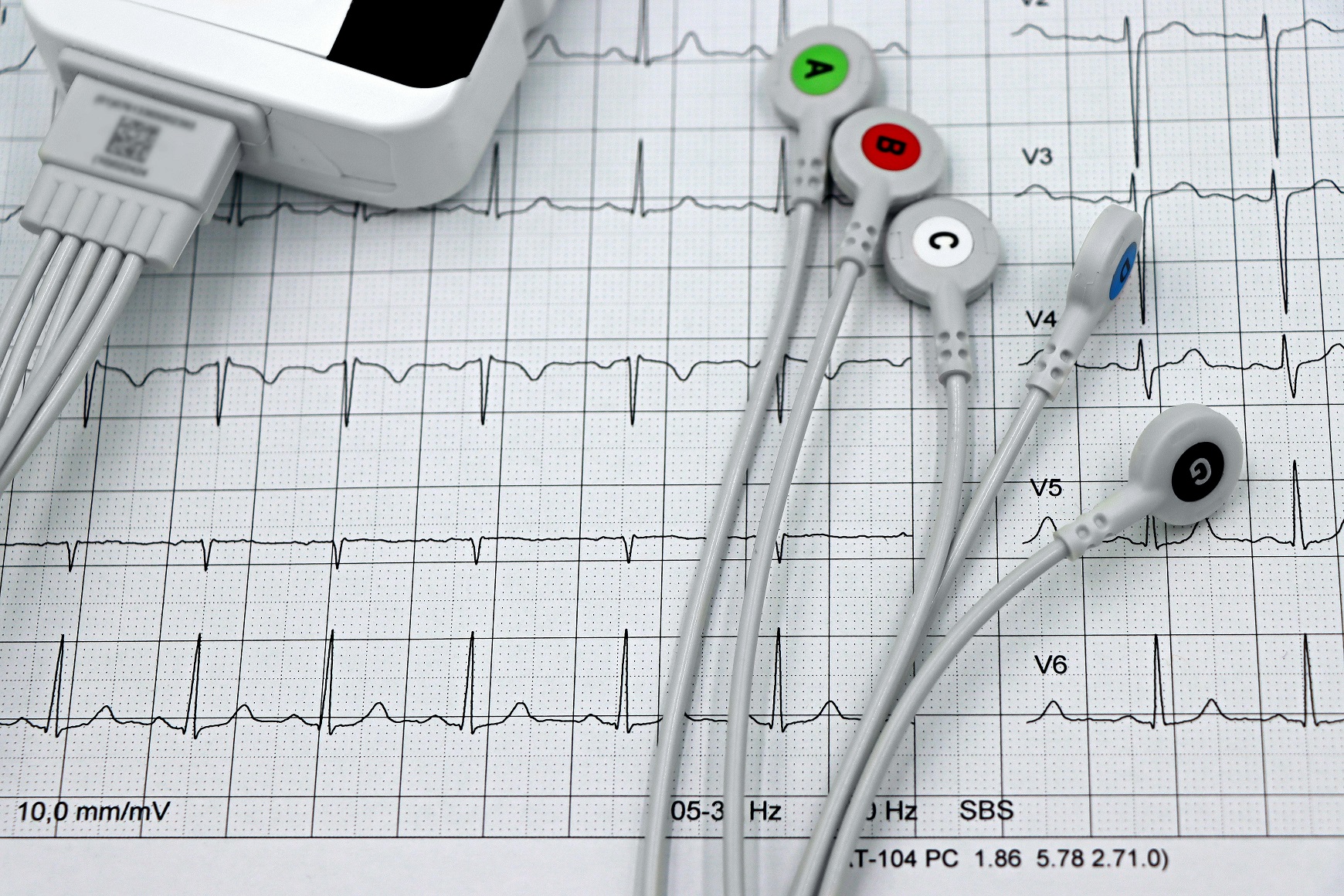
The interventional radiologist or cardiologist will make a small incision and then the doctor inserts a thin, flexible tube called a catheter into a blood vessel at the catheter site chosen. Using X-ray imaging for guidance, they carefully navigate the catheter to the targeted area.
A contrast material, or dye, is injected through the catheter to make the blood vessels visible on the X-ray images. You may feel a brief warmth or flushing sensation as the doctor injects the dye and it circulates.
As the dye moves through your vessels, a series of detailed X-ray images is taken. These images help identify blocked arteries, narrowing of vessels, or other abnormalities.
Once imaging is complete, the catheter is removed, and pressure is applied to the insertion site to prevent bleeding. In some cases, a small closure device may be used. You’ll then be monitored in a recovery area for several hours. Heavy lifting should be avoided for a week following the procedure.
While angiography and angioplasty are related procedures, they serve distinct purposes and are performed differently. Understanding these differences can help distinguish their roles in diagnosing and treating cardiovascular disease.
Angiography involves taking detailed X-ray images of blood vessels using a contrast dye. It helps identify problems like blockages, narrowing, or abnormalities in the blood vessels. Distinct types of angiograms include:
On the other hand, angioplasty is a treatment procedure aimed at opening blocked or narrowed blood vessels. It often follows an angiogram if significant blockages are found.
Coronary angiography is primarily a diagnostic tool where a catheter is inserted into a blood vessel to inject contrast dye, allowing doctors to visualize the vessels and pinpoint issues. The procedure stops at diagnosis.
After identifying a blockage, an angioplasty procedure may be performed. A small inflatable balloon at the end of a catheter is expanded at the site of a blockage to widen the artery. Sometimes, a stent (a small mesh tube) is placed to keep the artery open.
Angiography is considered minimally invasive. It is primarily diagnostic and involves no direct intervention in the blood vessel structure. Angioplasty is a therapeutic procedure and involves physical manipulation of the artery, making it more invasive than angiography. However, it is still considered minimally invasive when compared to more complicated surgeries like open surgery on the heart muscle or a bypass operation.
Angiograms provide a clear picture of vascular health but do not directly resolve blockages or treat conditions. It often informs further treatment decisions. Meanwhile, angioplasty directly addresses the issue, improving blood flow and potentially alleviating symptoms like chest pain (angina) or reducing the risk of heart attack.
Think of angiography like mapping out the problem, while angioplasty is taking immediate action to fix it. Both procedures often work hand-in-hand, with angiography paving the way for effective treatment through angioplasty or other medical interventions.
An angiogram is a vital diagnostic tool that helps physicians assess the health of blood vessels and identify underlying conditions. It’s typically recommended when symptoms or medical tests suggest problems with blood flow. Let’s examine common circumstances in which a doctor might recommend an angiogram.
Persistent or severe chest pain, especially if it worsens with activity, may indicate coronary artery disease or blockages. Difficulty breathing or shortness of breath without a clear cause may signal reduced blood flow to the heart or lungs. Pain in the arms, neck, jaw, or back could also be signs of reduced blood supply to the heart. Leg pain or cramping (claudication) while walking may suggest vascular conditions like peripheral artery disease (PAD).
If a treadmill or pharmacological stress test shows signs of reduced blood flow to the heart, an angiogram may be performed to identify potential blockages. Echocardiograms or CT scans can also return suspicious findings that might require an angiogram for confirmation.
Certain medical conditions can be accurately diagnosed or informed through the use of an angiogram.
While angiograms are generally safe and widely used, they do carry some risks, as with any medical procedure. While the following risks exist, angiograms are crucial for diagnosing and managing life-threatening conditions, and their benefits often outweigh potential complications. Understanding these potential risks can help patients make informed decisions and discuss concerns with their healthcare providers.
Some swelling, bruising, or minor bleeding where the catheter was inserted (usually the groin, arm, or wrist) is common and typically resolves quickly. The catheter’s insertion and movement can occasionally damage the blood vessel, leading to complications like arterial dissection (a tear in the artery) or hematoma (a collection of blood outside the vessel).
Rarely, the catheter can cause small blood clots to form, which may dislodge and lead to blockages elsewhere in the body.
The iodine-based contrast material used during the procedure can cause mild allergic reactions, such as itchy skin, rash, or nausea. Severe reactions are rare but can include difficulty breathing or anaphylaxis. Patients with a history of iodine or shellfish allergies should inform their doctor beforehand.
Though rare, there is a slight risk of infection at the catheter insertion site. Proper sterilization techniques reduce this risk significantly.
During an angiogram, a clot or debris may dislodge, potentially causing a heart attack or stroke, though this is extremely uncommon. The procedure may occasionally trigger arrhythmias (abnormal heart rhythms), which typically resolve quickly.
The angiogram stands as a shining beacon in clinical practice guidelines for quickly and accurately imaging circulatory issues. It’s a diagnostic tool that can help healthcare providers identify coronary heart disease, aneurysm, or stroke. While there are risks, many doctors value the insights it provides and consider it a vital tool for patient care.
Browse top-rated specialists and book an appointment with ease.
Find a DoctorSubscribe to our newsletter for expert health insights, the latest treatments, and tips from top specialists—delivered straight to your inbox.


0 comments